Medical ultrasound
Imaging technique for the examination of organic tissue
Medical ultrasound is an imaging technique for the examination of organic tissue. It is the most commonly used imaging technique in medicine.
Within the field of urology, ultrasound is well suited for the diagnosis of pathological changes to the kidneys, urinary bladder, testicles and epididymis. The adrenal glands and ureters are usually not detectable using ultrasound. Gas-containing organs, such as the lungs, intestines or bones, are also generally difficult to examine using ultrasound.
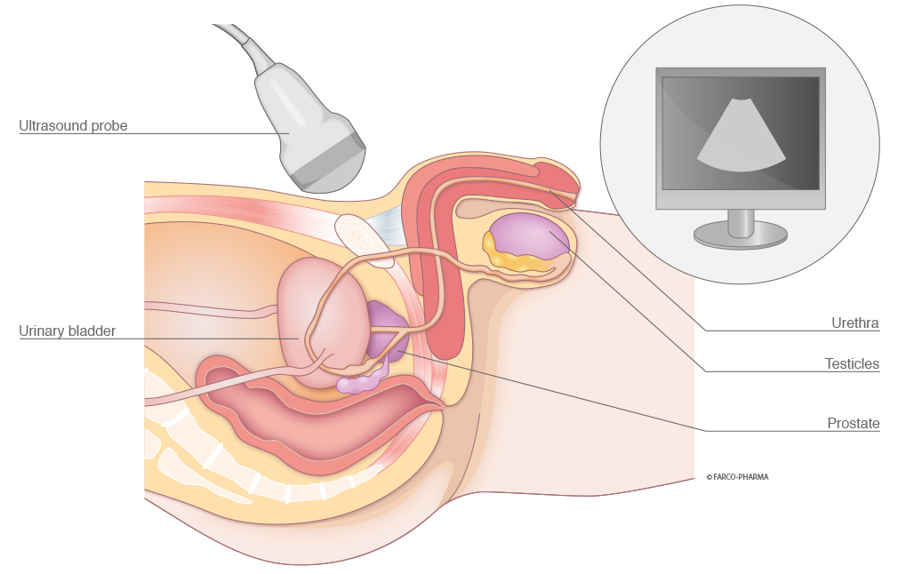
The prostate holds a special position. If necessary, the prostate is examined using transrectal ultrasound with a probe that is inserted into the anus.
Sequence of the examination
Ultrasound examinations involve the examining physician placing an ultrasound probe on the skin of the region of the organ to be examined. The ultrasound probe is coated with a contact lubricant in advance, e.g. InstillaGel® Lubri, in order to enable the examination. The examination can last several minutes because organs are viewed in individual layers. With the aid of soundwaves, images of the inside of the body are produced that the physician is able to view on a monitor connected to the ultrasound probe. This enables the direct identification of potential changes to organs and tissue during the examination and the photo-documentation of the findings (sonogram).
Risk-free and painless examination
A major advantage of ultrasound over radiography is the innocuousness of the soundwaves in use. Due to the fact that no harmful rays are emitted, ultrasound examinations are risk-free and painless. As a result, it is also possible to perform these examinations on pregnant women.