Transrectal ultrasound (TRUS)
What is TRUS?
Transrectal ultrasound (TRUS) is an imaging technique that is used to diagnose changes in the prostate and seminal vesicles. TRUS is a form of endoscopic ultrasound, a type of ultrasound that involves the insertion of a transducer into the body often with the aid of an endoscope or a probe.
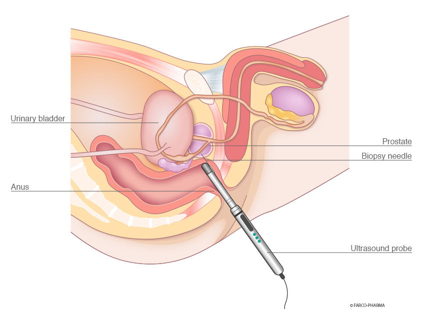
For this examination, the transducer is mounted to an ultrasound probe that is approximately the size of a finger. The probe is then inserted into the rectum via the anus and positioned near the prostate. The prostate is located immediately upstream from the rectum and is especially easy to examine using TRUS because the ultrasound signal is only dampened by a few interstitial tissues. Transrectal ultrasound provides precise images of the prostate, seminal vesicles and their surroundings.
Fields of application for transrectal ultrasound
Transrectal ultrasound is used to clarify cases of enlarged prostate, abnormal palpation findings and elevated PSA values (prostate-specific antigen) in the blood. Another important field of application for transrectal ultrasound is the visualisation of tissue collection from the prostate (prostate punch biopsy).
TRUS enables a more precise assessment of the appearance of the prostate and the seminal vesicles because this examination method provides precise images of the prostate, seminal vesicles, and their surroundings and a differentiated representation of the dimension, shape, delimitation, and internal structure (zones) of these organs, as well as enabling the identification of abnormal areas. Based on the measured dimensions of the prostate, its volume is able to be calculated using software. This enables the detection of any changes in the shape or boundaries of the prostate. In the case of suspected cancer-related changes, transrectal ultrasound is used to collect samples (biopsies) from so-called hypodense areas. If prostate cancer is present, then TRUS is able to clarify the tumour stage prior to surgery. This involves the determination of whether the tumour has extended beyond the prostatic capsule or infiltrated the seminal vesicles.
Transrectal ultrasound is also used to detect the stasis of secretions, prostatic calcifications and seminal vesicle wall thickening.
Sequence of the examination
The examination is performed with the patient in the supine or lithotomy position. It is normally not necessary to prepare the colon, but the rectal ampulla should be emptied.
As in the case of digital rectal examinations (DRE), transrectal ultrasound is also painless provided that there are no painful changes in the rectal region. The examination lasts a few minutes and is an outpatient procedure.
A sterile lubricant with local anaesthetic and disinfecting properties, such as Instillagel®, is instilled into the anus and rectum prior to the examination. Following an exposure time of 5 - 10 min, the ultrasound probe is inserted into the rectum like a finger. By moving the probe slightly, the physician establishes the areas to be examined that are visible on the monitor. The findings on the monitor are able to be photo-documented during the examination. Pain or complications are not to be expected by patients during or after the examination.